

CBCT Interpretation for Implant Planning: A Practical Starting Point
ConfiDental Education Center
Programme team
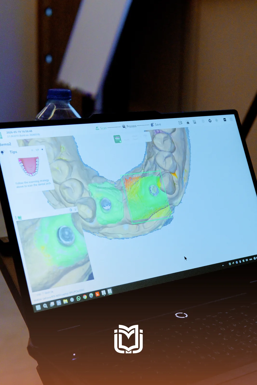
A panoramic radiograph tells you a tooth is missing. It does not tell you whether you can safely put an implant where it used to be.
That gap — between two-dimensional imaging and a three-dimensional surgical decision — is where a lot of early implant cases go wrong. Cone beam computed tomography closes it, but only if you know what you are looking at.
This is a practical starting point for reading a CBCT scan with implant placement in mind. It is not a substitute for structured training, but it will tell you what questions the scan is supposed to answer.
Start with the restoration, not the bone
The most common planning error is opening the scan and looking for bone. That is backwards.
Prosthetically driven implantology means you decide where the crown needs to be first, then ask whether the bone will let you put an implant under it. Plan from available bone instead and you end up with implants in positions that were convenient to place and are difficult to restore properly.
Plan the crown. Then ask the bone whether it agrees.
In practice that means a radiographic guide, or at minimum a clear model of the final restoration, before you measure anything.
The four measurements that matter
Once you know where the restoration goes, the scan needs to answer four questions.
What to measure, and why
- Available height — crest to the nearest vital structure. Decides implant length and whether augmentation enters the conversation.
- Available width — measured at the depth the implant body will sit, not at the crest. Ridges narrow apically more often than people expect.
- Bone quality — the cortical and cancellous pattern informs your expectation of primary stability and your drilling protocol.
- Proximity to vital structures — inferior alveolar canal, mental foramen, maxillary sinus, nasal floor, adjacent roots.
Vital structures: where people get caught out
The inferior alveolar canal is the structure everyone checks. The ones that catch people out are the ones they do not.
The anterior loop of the mental nerve can extend mesial to the mental foramen, and it is invisible on a panoramic. Treating the interforaminal region as an automatic safe zone is a well-recognised route to a neurosensory complication.
In the maxilla, the sinus floor is rarely flat. Septa are common, and a scan reviewed only in one plane can miss one sitting directly in your planned osteotomy.
Adjacent root proximity matters especially in the aesthetic zone, where an angulation that looks fine on a panoramic turns out to be very close to a neighbouring root on the axial slice.
Read the scan in all three planes, every time
A CBCT is a volume, not an image. Each plane answers a different question.
| Plane | What it shows best |
|---|---|
| Cross-sectional | Ridge width, buccolingual angulation, undercuts, sinus floor |
| Panoramic (reconstructed) | Overall anatomy, mesiodistal spacing, canal course |
| Axial | Root proximity, ridge shape at a given depth, arch form |
| 3D reconstruction | Communicating the plan — useful for the patient, not for measuring |
The 3D reconstruction looks the most impressive and is the least useful clinically. Measure on the slices.
Common interpretation mistakes
Five things worth checking in your own planning
- Measuring ridge width at the crest instead of at implant depth.
- Trusting an automatic nerve trace without verifying it slice by slice.
- Treating the interforaminal region as automatically safe.
- Planning from bone availability rather than from the restoration.
- Not reviewing the volume for pathology unrelated to the implant site.
Where a scan stops being enough
A CBCT describes hard tissue. It tells you very little about soft tissue thickness, keratinised tissue width, or biotype — all of which affect the aesthetic result and long-term stability.
It also cannot tell you whether the patient is a good candidate. Smoking, glycaemic control, parafunction and periodontal status sit outside the scan and inside the case selection decision.
The scan narrows your options. It does not make the decision.
Learning to do this reliably
Reading CBCT for implant planning improves with supervised repetition on real cases, not with reading. The Clinical Implant Program opens with CBCT interpretation, planning software workflow and prosthetically driven planning before any surgical content — deliberately, because everything downstream depends on it.